in this Blog, we will discuss acute cholecystitis.
cholecystitis is the inflammation of the gallbladder most commonly caused by gallstones. it develops in six to eleven 11 of patients with symptomatic gallstones. it is more common in women than men and has a higher prevalence in western caucasian Hispanic and Native American patients.
so what causes cholecystitis?
the pathophysiology of cholecystitis and acalculous cholecystitis can be calculus with a stone or a calculus without a stone which is less common in only about five to ten percent of cases, it often occurs in the setting of cystic duct obstruction with a stone leading to increased internal pressure which releases inflammatory mediators which leads to ischemia of the gallbladder wall and can ultimately lead to necrosis.
let's look at risk factors.
the risk factors for stone formation include the female sex having a family history, pregnancy, diabetes mellitus, dyslipidemia, and obesity. the use of the antibiotic ceftriaxone can cause sludge and lead to calculus cholecystitis just as prolonged use of total parental nutrition or TPN can.
let's look at the signs and symptoms of acute cholecystitis.
characteristically acute cholecystitis pain occurs in the epigastric region and most commonly in the right upper quadrant region,
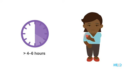
it is steady and severe and is typically prolonged lasting more than four to six hours the patient may present with pain that radiates to the right shoulder or back due to peritoneal inflammation of the diaphragm.
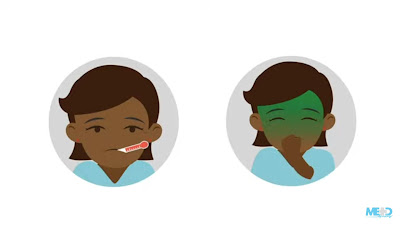
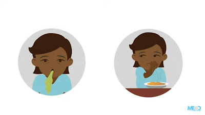
they may have associated complaints including fever, nausea, vomiting, and anorexia. there is often a history of fatty food ingestion one hour or more before the initial onset of pain.
now let's work up a patient with acute cholecystitis.
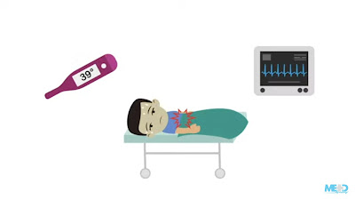
patients with acute cholecystitis are usually ill-appearing on inspection they may also have jaundice check not only on their skin but their eyes for scleral icterus patients will often lie still on the examining table because cholecystitis is associated with local parietal peritoneal inflammation that is aggravated by movement their vital signs may reveal fever and tachycardia the abdominal exam will usually show right upper quadrant pain and possibly even epigastric pain the patient may demonstrate voluntary or involuntary guarding to palpation as well cholecystitis can cause a positive murphy sign which is a sign of peritoneal inflammation for details on performing a full abdominal exam please see our men mastery course abdominal examination essentials.so what might you expect to see in the lab results of a patient with cholecystitis?
patients with cholecystitis typically have a leukocytosis and a left shift on their complete blood count the liver function tests are important in these patients a liver function test will report not only the total bilirubin but will specify the portion that is conjugated or direct versus unconjugated or indirect. patients with cholecystitis may also have elevations in their serum aminotransferases which are the AST and ALT in addition to a slight elevation in their serum amylase or alkaline phosphatase levels.
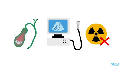
so what about imaging ultrasound is a diagnostic modality of choice for suspected cholecystitis.
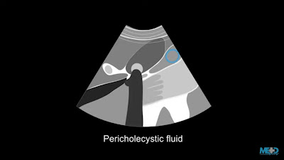
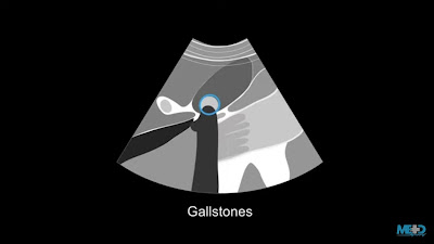
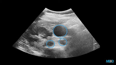
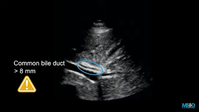
it should be the first imaging modality to use and often the only one needed to make the diagnosis. ultrasound is both sensitive and specific for evaluating gallbladder disease it is also minimally invasive without the radiation risk. the finding on ultrasound that indicates cholecystitis or acalculous cholecystitis are gallbladder wall thickening equal to and greater than four millimeters of pericholecystic fluid and wall edema sometimes called a double wall sign and gallstones which will be seen as acoustic shadows here is an example of a patient's gallbladder ultrasound in the transverse plane you can see cholecystitis icd10 acoustic shadows representing gallstones the common bile duct size can be very well seen with ultrasound a diameter greater than 8 millimeters or 0.8 centimeters can be concerning for obstruction of the common bile duct and usually warrants further investigation of the common bile duct for example with magnetic resonance cholangitis cryptography or mrcp. the common bile duct generally increases in size by one millimeter for every decade of life.
so someone who is 80 years old or more significant may have a larger common bile duct that is not pathologic a couple other things that can enlarge the common bile duct are previous cholecystectomy as well as recent instrumentation such as that done with endoscopic retrograde cholangitis cryptography or ERCP.
for details on performing an ultrasound of the gallbladder and bile duct please check out menmastery's point of care ultrasound essentials course.
let's discuss the treatment for acute cholecystitis.
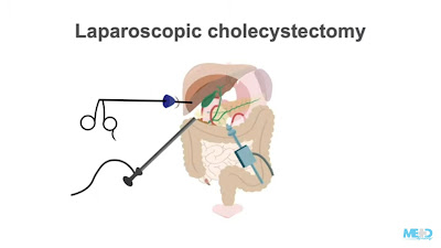
medical treatment options are usually temporary and will not treat the underlying disease because the gallbladder is the source of stones and must be treated by removal it is only a matter of time before surgery is needed. laparoscopic cholecystectomy is a gold standard for the treatment of acute cholecystitis and is a preferred form of cholecystectomy. it can also be robotically done which is also a well-accepted minimally invasive approach. laparoscopic cholecystectomy has the benefits of faster recovery and returns to work with smaller wounds and scars and a lower risk of complications. if a patient is too ill to undergo surgery but has an acute closed status that needs to be immediately treated. a percutaneous cholecystectomy tube which is often placed by interventional radiology and can be used to have controlled drainage of the gallbladder in the acute setting is placed ultimately it can be removed and cholecystectomy performed once the surgical contraindications are mitigated waiting usually six to eight weeks or longer to proceed.







If you have any doubts, let me know ...